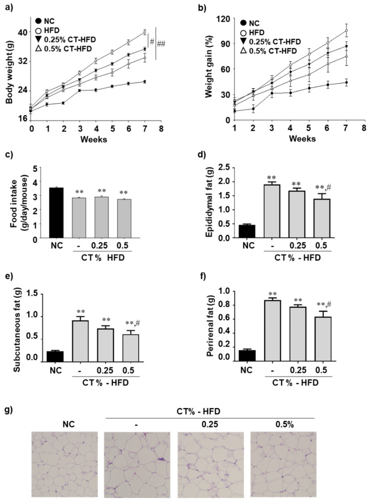
Figure 1.
Effect of CT treatment on body weight, fatty tissue mass, and adipose tissue histopathology. One week after acclimatization, C57BL/6J mice were fed HFD mixed with 0.25% and 0.5% CT for 7 weeks. (a,b) Body weights and weight gain were measured (n = 8–9); (c) Food intake was monitored weekly; (d–f) Epididymal, subcutaneous, and perirenal adipose tissues were weighted; (g) H&E staining was performed on epididymal adipose tissue sections. NC: normal chow; (-): HFD; CT-HFD; CT treated HFD. Values are mean ± SE. AVOVA p-value: ** <0.01 vs. NC group; ## <0.01, # <0.05, vs. HFD-fed group.

