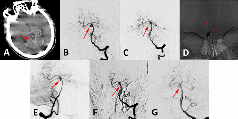
Fig. 2.
A ruptured basilar trunk aneurysm with a daughter sac was treated with LVIS-assisted coiling, and computed tomography confirmed subarachnoid hemorrhage (a arrow). Compared with the anteroposterior position in the preoperative angiographic images (b arrow), the aneurysm was completely occluded after treatment in the immediately post-procedure angiograph (c arrow). The LVIS stent was placed across the neck of the aneurysm followed by embolization with coils (d). At the 6-month follow-up angiography, the aneurysm had recanalized (e arrow). Repeat coiling was performed, and the residual aneurysm neck was occluded completely (f arrow). At the 7-month follow-up angiography, the aneurysm was stable and completely occluded (g arrow)

