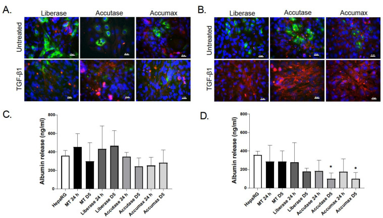
Figure 3.
Cellular characteristics and functionality of cells obtained by dissociation of MTs. MTs were dissociated at day 3 (A) or 9 (B) using Accutase, Liberase or Accumax. Isolated cells were seeded directly into a 96-well and allowed to attach for 3 days. Replated cells were treated with TGF-β1 (1 ng/mL) for 48 h. Activation of hTERT-HSCs was determined by α-SMA immunostaining and functionality of HepaRGs by albumin immunostaining. Staining for albumin is in green, α-SMA in red and DAPI in blue. Photomicrographs are taken at 40× magnification scale bar is 20 µm. Albumin secretion of cells dissociated at day 3 (C) and day 9 (D) was determined 24 h and 5 days after re-plating using an ELISA. Data are expressed as albumin concentration ± SD of n = 6–9. Statistical analysis using unpaired student’s t-test comparing dissociated cells to MT for each time point; *: p ≤ 0.05.

