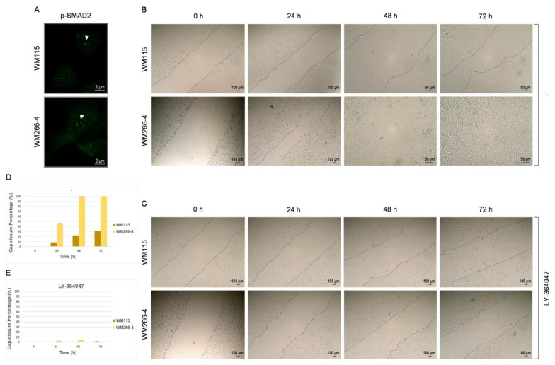
Figure 3.
In vitro migration of WM115 and WM266-4 human melanoma cells depends on TGF-β/SMAD2 signaling axis. (A) Immunofluorescence patterns of (activated) “p-SMAD2” protein in WM115 (primary) and WM266-4 (metastatic) melanoma cells. “p”: Phosphorylation. Arrowheads: “p-SMAD2”-positive nuclear “specks”. Scale bars: 2 μm. (B,C) Light micrographs of wound healing (scratch wound) assays, examining the motility/migration capacities of WM115 and WM266-4 melanoma cells, in the absence (B) or presence (C) of the TGF-β signaling specific inhibitor LY-364947 (100 μM), for a time-frame of 0–72 h. (B) Scale bars: 50 or 100 μm. (C) Scale bars: 100 μm. Dash lines denote the melanoma cell migration borders. (D,E) Quantification, in bar-chart format, of in vitro motility/migration activities, as demonstrated by gap-closure percentages (%) (mean values) in wound healing assays (B,C) of WM115 (primary) and WM266-4 (metastatic) human melanoma cells in the absence (B,D) or presence (C,E) of the LY-364947 inhibitor (0–72 h).

