Figure 5.
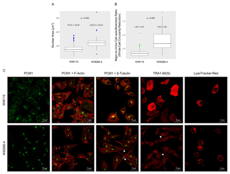
IMC program implementation in BRAFV600D melanoma cells requires re-modeling of cytoskeleton architecture. (A,B) (Bio-)metrical features of WM115 (primary) and WM266-4 (metastatic) melanoma cell size and morphology/geometry using vimentin-derived patterning (Figure 2A). (A) “Nuclear Area” (size) (μm2). (B) “Major-to-minor cell-axis/dimension ratio” (whole cell circularity reduction). Spindle-like (elongated) “geometrical” shapes fit the fibroblast-like phenotypes of WM266-4 metastatic melanoma cells. (C) Single immunofluorescence profiles of “PCM1” and “TRA1-60(S) (PODXL)” proteins in WM115 (primary) and WM266-4 (metastatic) melanoma cells. Double (immuno)fluorescence patterns of “PCM1 and F-Actin”, and “PCM1 and β-Tubulin” proteins, with “β-Tubulin” (red) and “PCM1” (green) being detected via antibody-based fluorescence, and “F-Actin” being identified by rhodamine-phalloidin staining (red) in WM115 (primary) and WM266-4 (metastatic) melanoma cells. The yellow color is derived from the merger of green and red colors. Fluorescence profiles of lysosomes distribution (red) in WM115 (primary) and WM266-4 (metastatic) melanoma cells. Arrowheads: “β-Tubulin” (MT)-positive, lengthy, Invadopodia, and “TRA1-60(S)”-positive Invadopodia. “F”: Filamentous. “β”: beta. Scale bars: 2 or 5 μm.

