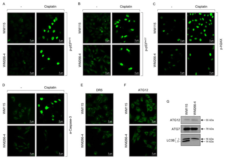
Figure 8.
Programmed cell death sub-routines in BRAFV600D-positive primary and metastatic melanoma cells. (A) Immunofluorescence patterns of the “p-p53Ser15” protein in WM115 (primary) and WM266-4 (metastatic) melanoma cells, in the absence (−) or presence of cisplatin (50 μg/mL, for 24 h) chemotherapeutic agent. “p”: Phosphorylation. “Ser”: Serine. Scale bars: 5 μm. (B) Immunofluorescence profiles of the “p-p53Ser37” protein in WM115 (primary) and WM266-4 (metastatic) melanoma cells, in the absence (−) or presence of cisplatin (50 μg/mL, for 24 h). “p”: Phosphorylation. “Ser”: Serine. Scale bars: 5 μm. (C) Immunofluorescence patterns of the “p-H2AX” protein in WM115 (primary) and WM266-4 (metastatic) melanoma cells, in the absence (−) or presence of cisplatin (50 μg/mL, for 24 h). “p”: Phosphorylation. Scale bars: 5 μm. (D) Immunofluorescence profiles of the (cleaved/activated) “a-caspase-3” protein in WM115 (primary) and WM266-4 (metastatic) melanoma cells, in the absence (-) or presence of cisplatin (50 μg/mL, for 24 h). “a”: Activated. Scale bars: 5 μm. (E) Immunofluorescence patterns of the “DR5” transmembrane death receptor in WM115 (primary) and WM266-4 (metastatic) melanoma cells. Scale bars: 5 μm. (F) Immunofluorescence profiles of the “ATG12” autophagy protein in WM115 (primary) and WM266-4 (metastatic) melanoma cells. Scale bars: 5 μm. (G) Western blotting-mediated examination of the “ATG12”, “ATG7”, and “LC3B (I/II)” protein expression profiles in WM115 (primary) and WM266-4 (metastatic) BRAFV600D-dependent human melanoma cells. Molecular weights of the herein identified autophagy proteins are denoted by numbers at the right side of each respective panel. (G) Protein quantification values (in bar-chart format) are shown in Figure S1.

