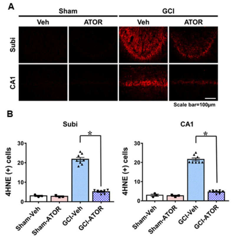
Figure 2.
Atorvastatin reduces ischemia-induced oxidative stress. Neuronal oxidative stress was detected through 4HNE (red color) staining in CA1 and subiculum 1 week after ischemia. The sham-operated groups showed a weak red fluorescence in hippocampal CA1 and subiculum. The GCI-ATOR group showed a weaker 4HNE fluorescence intensity compared with the GCI-Veh group (A). Scatter dot plot bar graph represents the quantified 4HNE intensity in each region (B). Data are mean ± S.E.M.; n = 3 for each sham group, n = 8 for each ischemia group. * Significantly different from the vehicle-treated group, * p < 0.05.

