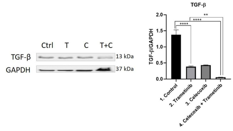
Figure 11.
Western blot analysis of TGF-β. Protein expression of TGF-β in BJ + SK-MEL-28 co-culture cells. WB bands were analyzed by densitometry and results were normalized to GAPDH. The left panel illustrates WB bands (Ctrl = control, T = trametinib, C = celecoxib, T + C = trametinib + celecoxib), while right panels illustrate the quantitative analysis of WB results (1 = control, 2 = trametinib, 3 = celecoxib, 4 = celecoxib + trametinib). ** p < 0.01, **** p < 0.0001. Each bar represents mean ± standard deviation (n = 3).

