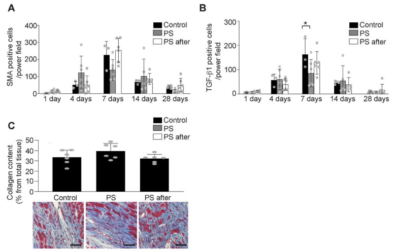
Figure 5.
The effect of phosphatidylserine on remodeling after AMI. (A) The number of myofibroblasts (α-SMA positive cells) in the infarct area increased following permanent coronary ligation with no difference between the control or PS treated groups (n = 5–6/group, Two-way ANOVA, Bonferroni’s multiple comparison Test, Values ± SD). (B) TGF-β1 expression is less pronounced after phosphatidylserine treatment (n = 5–6/group, Two-way ANOVA, Bonferroni’s multiple comparison Test, * p < 0.05 vs. Control, Values ± SD). (C) There was no difference in myocardial collagen content (blue, Gomori staining) expressed as % from infarcted area at 28 days after AMI between control (Control) and phosphatidylserine-treated mice (PS, PS after) (n = 5–6/group, One-way ANOVA, Turkey’s multiple comparison Test, Values ± SD). Representative images of Gomori staining (lower panel, scale bar 50 µm).

