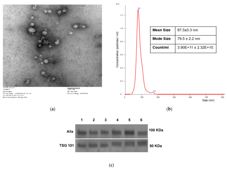
Figure 1.
Characteristics of sEV isolated from NSCLC plasma. (a) Transmission electron microscopy of isolated NSCLC exosomes. (b) Size and concentration of NSCLC exosomes as determined by tunable resistive sensing (TRPS). (c) TSG101 and ALIX detection by Western blots in sEV isolated from plasma of several different patients with NSCLC. Each lane was loaded with 10 µg sEV protein present in fraction #4.

