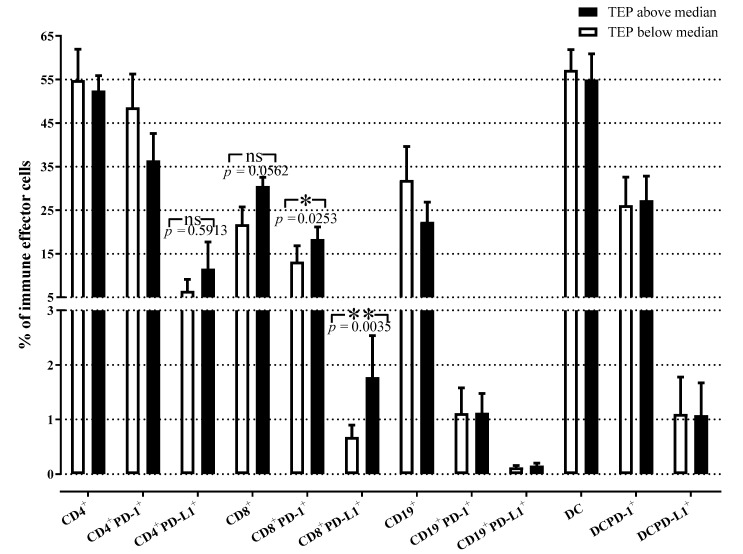
Figure 4.
Relationship between TEP levels and peripheral immune cells in patients with NSCLC. Percentages of different effector immune cell subpopulations in stage IV patients are related to the TEP levels at baseline. The percentages of CD8+PD-1+ and CD8+PD-L1+T cells were significantly increased in patients with high TEP levels as compared to patients with low TEP levels. The bars denote mean values ± SEM. Groups were compared by Mann–Whitney U test (TEP above vs. below median for CD4+PD-L1+, CD8+PD-L1+, CD19+PD-L1+, DCPD-L1+) and unpaired t-test (TEP above vs. below median for CD4+, CD4+PD-1+, CD8+, CD8+PD-1+, CD19+, CD19+PD-1+, DC, DCPD-1+; open bars = TEP below the median, closed bars = TEP above the median; TEP = total sEV protein; *, p < 0.05; **, p < 0.01).

