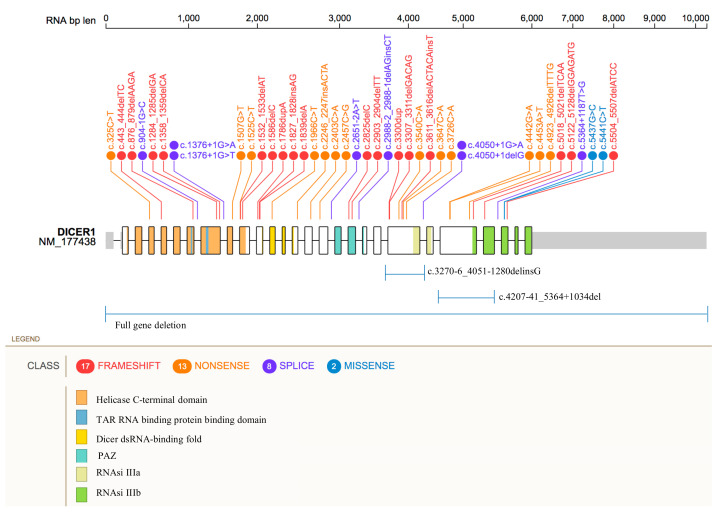
Figure 1.
Distribution of DICER1 germline pathogenic variants. Figure 1 shows the location of the genetic variants along the DICER1 gene. SNPs and indels mutations are reported as frameshift (red), nonsense (orange), splice-site (purple), and missense (blue) variants. Large deletions are shown below. Protein domains are represented by colored areas.

