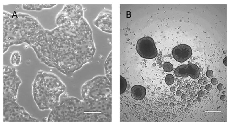
Figure 3.
Morphology of 82.3 cells. (A) Cells were able to grow in monolayer and exhibited epithelial morphology (4× magnification); (B) representative images of cholangiospheres of 82.3. Cells were seeded in ultra-low attachment plate and stem cell-serum-free medium. Sphere formation was monitored on days 7, 10, and 14 after seeding (4× magnification).

