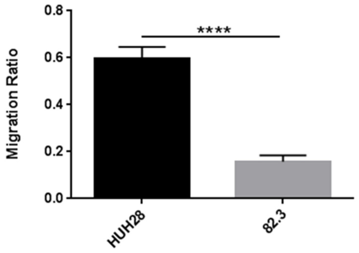
Figure 6.
Transwell migration assay on 82.3 and HUH28 cells. Cells were seeded on the surface of the migration transwell chamber and separated by a porous membrane. After 48 h of incubation, the membranes were fixed with methanol and stained with crystal violet. The area of the cells that invaded the membrane was calculated using the ImageJ 2 software; five different fields were evaluated. Migration is expressed as the ratio of the mean ± SEM of the area of migrated cells to the area of the same number of cells plated (control). **** p < 0.00001.

