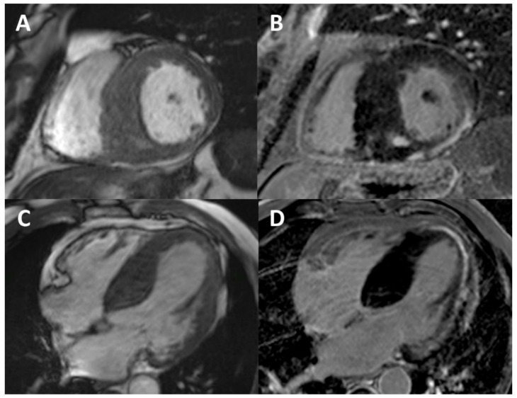
Figure 4.
Cardiac MRI (3.0 Tesla) in a Fabry patient with advanced cardiomyopathy. Cine (balanced steady-state free precession sequence) images at the basal LV short-axis slice (A) and four-chamber view (C) showing massive and asymmetrical LVH (maximal thickness 30. mm at the septum) with thinning of the posterior wall (2 mm). LGE at the basal LV short-axis slice (B) and four-chamber view (D) showing fibrosis of the inferior and inferolateral walls and apex and focal fibrosis in the septum, where the LVH is more pronounced.

