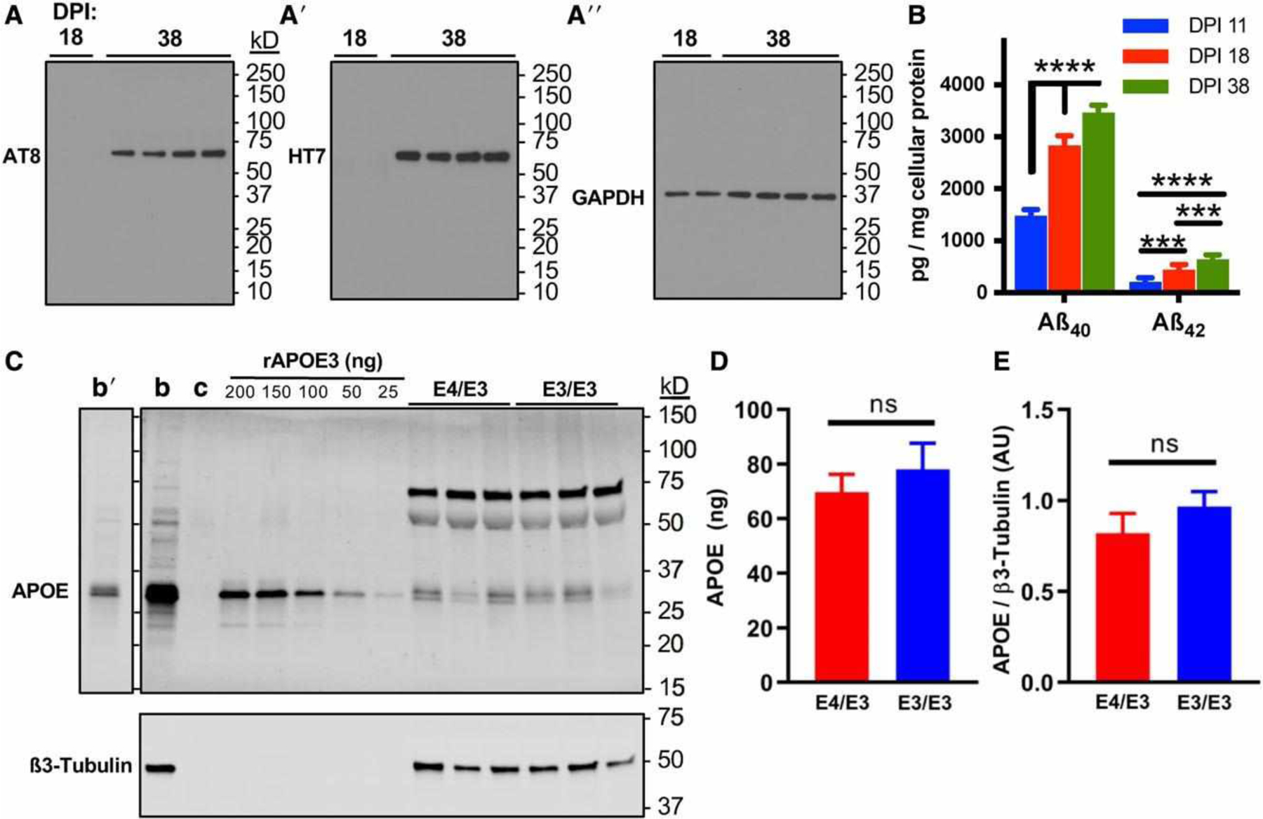
FIGURE 3:

Forebrain excitatory neurons express p-tau, Aß, and apolipoprotein E (APOE). (A) Immunoblotting of p-tau detected by AT8, total tau detected by HT7, and glyceraldehyde-3-phosphate dehydrogenase (GAPDH) during the maturation of neuronal cultures at 18 and 38 days postinduction (DPI). Blots were stripped and probed sequentially. (B) Aß40 and Aß42 peptides were detected by peptide-specific immunoassay in the cell culture supernatant of developing and mature neurons by 11 DPI, and secretion increased over time until plateauing by 38 DPI. There were n = 3 independent differentiations. Each data point is the average of 3 measurements per differentiation. The statistical test was a 2-way repeated measures analysis of variance followed by Tukey range test: ***p < 0.001, ****p < 0.0001. (C) Isogenic E4/E3 and E3/E3 neurons at 38 DPI express APOE. Representative immunoblot depicts APOE with ß3-tubulin to control for neuronal protein loading. b = positive control human adult brain lysate; c = negative control lysate of HEK293 cells transfected with an empty vector. Recombinant APOE3 (rAPOE3) purified from HEK293 cells was serially diluted to generate a standard curve to quantify the expression of APOE in lysates from E4/E3 and E3/E3 neurons. Human brain lysates are also depicted at a shorter exposure time (b0). (D, E) Absolute (D) and relative (E) quantitation of immunoblotting experiments in C. There were n = 6 independent differentiations. The statistical test was 2-tailed unpaired Student t test: ns = not significant.
