
FIGURE 6:
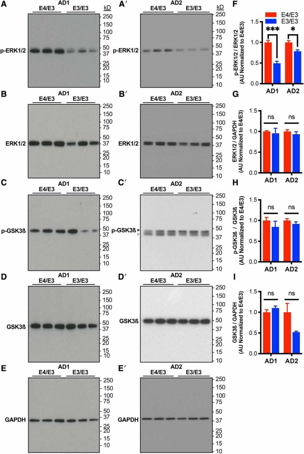
Neurons expressing E4 exhibit increased phosphoactivation of ERK1/2 but not GSK-3ß. (A–E) Representative immunoblotting of lysates of isogenic E4/E3 and E3/E3 neurons from AD1 and AD2 with antibodies directed against phosphorylated or total ERK1/2 and GSK3ß. Glyceraldehyde-3-phosphate dehydrogenase (GAPDH) was used as a control for protein loading. Blots were stripped and probed sequentially. Open arrowhead indicates residual ERK1/2 immunoreactivity not used for quantitation. (F–I) Quantitation of phosphokinase/total kinase ratio and total kinase expression from A–E. There were n = 6 independent differentiations per genotype and patient background. Measures are normalized to the E4/E3 genotype within each patient background. The statistical test was Student t test between isogenic E4/E3 and E3/E3 neuronal lysates with Sidak correction for multiple comparisons: *p < 0.05, ***p < 0.001; ns = not significant.