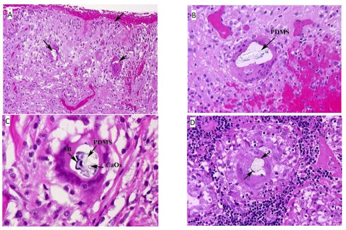
Figure 1.
(A) Fibrous capsule surrounding the implant, right. The surface of the tissue lies adjacent to the silicone breast implant and forms the “pseudosynnovium.” This pseudosynnovium is composed of amorphous eosinophilic material and mononuclear cells. Of note are several foreign body giant cells containing clear silicone gel. There is a chronic inflammatory infiltrate scattered among the fibroblasts, fibrous tissue, and collagen. (HE, 10×). (B) Fibrous capsule surrounding implant, right. A high-power view of the above tissue shows silicone gel within the giant cell that is embedded in fibrous tissue. There are also scattered chronic inflammatory cells, congested blood vessels, and surgical related hemorrhage. (HE, 100×). (C) Fibrous capsule surrounding implant, left. High power view of previous section. In the center of the multinucleated giant cell are fragments of Schaumann body (annotated on the image as SB), and refractile clear crystalline material consistent with calcium oxalate (annotated on the image as CaOx). (HE, 100×). (D) Axillary lymph node, left. There is clear refractile globular material in the center of the giant cell of the epithelioid granuloma consistent with silicone gel. In addition, fragments of a Schaumann body can be seen around the outer edge of the silicone gel. (HE, 100×). Arrows on the HE images, unless designated point towards areas of interest such as migrated PDMS.

