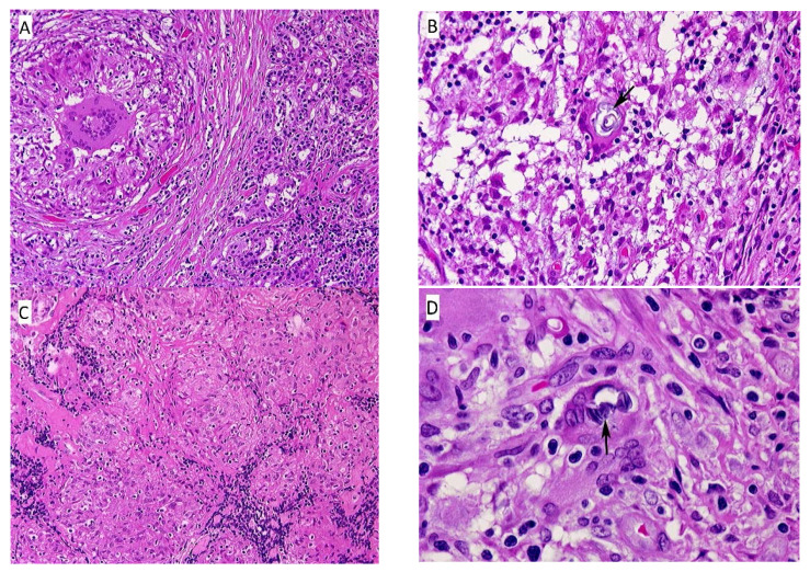
Figure 2.
(A) Eyelid nodule, left. This section contains both normal serous glands of the eyelid (right), and fibrous tissue surrounding a non-caseating granuloma with central foreign body giant. (HE, 10×); (B) Eyelid nodule, left. This section shows a non-caseating granuloma with a Schaumann body within the foreign body giant (center). (HE, 100×); (C) Lower leg nodule, left. There are aggregates of varying-sized epithelioid granulomas, fibrous tissue septae, and scattered lymphocytes. (HE, 10×); (D) Lower leg nodule, left. The section contains a non-caseating granuloma with scattered lymphocytes and several multinucleated giant cells in a fibrous tissue background. The giant cell in the center, indicated by the black arrow, contains two Schaumann bodies. (HE, 100×).

