
Figure 3.
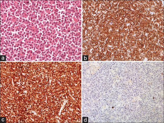
Histopathology. (a). Hematoxylin and eosin staining revealed diffuse proliferation of plasma cells with minimal pleomorphism, small number of large nulei and bi- or multinucleated cells. (H&E, ×200) (b). CD38+ (c). Immunohistochemical staining revealed kappa immunoglobulin light chains restriction. (d). Negative staining for lambda immunoglobulin light chains