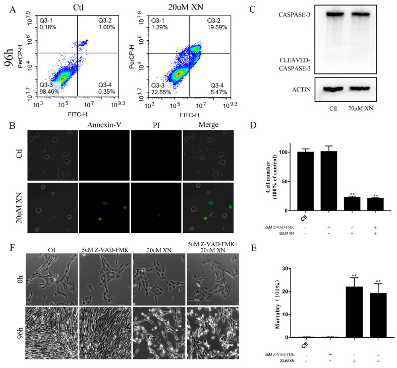
Figure 2.
XN-induced apoptosis of C6 cells. (A,B) C6 cell apoptosis evaluation by flow cytometry and fluorescence microscopy through Annexin-V/PI double staining after treatment with XN for 96 h (400× g magnification). (C) Cleaved caspase-3 was detected by Western blot (WB) after XN incubation for 96 h. (D–F) The C6 cells were pretreated with 5 uM inhibitor of caspase-3 Z-VAD-FMK for 1.5 h before the co-treatment with XN for another 96 h. The morphology changes were captured by a Leica Microsystems microscope (200× g magnification), and cell viability and cell death were measured by trypan blue staining. All data presented are the mean ± SEM from three independent experiments. ** p < 0.01 vs. control.

