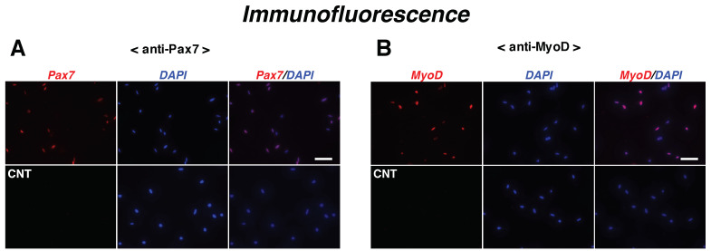
Figure 1.
Myogenic capability of satellite cells. (A,B) Satellite cell primary cultures isolated from the back, buttock, and upper hind limb were maintained for 24 h in F-10–20% FBS medium and evaluated for the ratio of cells expressing each myogenic protein. Immunofluorescence microscopy for Pax7 (panel A; red) or MyoD (panel B; red) and DAPI (blue) showing satellite cells and nuclei. CNT, control cells stained without primary antibodies. Scale bar in panels (A,B), 50 μm.

