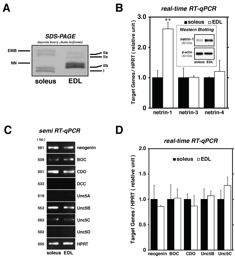
Figure 3.
Comparative analysis of netrin expression levels in EDL and soleus muscle-derived satellite cells. (panel A) Statues of soleus and EDL muscle; myofiber type composition of the skeletal muscle. MyHC isoforms (myofiber type; I [slow-twitch], IIa [intermediate], and IIx and IIb [fast-twitch]) were separated using SDS polyacrylamide gel electrophoresis (SDS-PAGE) of whole muscle samples (100 ng protein) and then visualized by silver staining. (panel B–D) Proliferated satellite cells from soleus and EDL muscles were maintained in differentiation medium until 72 h. (panel B and D) The cell lysates were evaluated for the mRNA expression of netrin-1, -3, and -4, neogenin, BOC, CDO, Unc5B, and Unc5C by real-time RT-qPCR standardized to HPRT expression. The bars depict the mean ± SEM of three independent cultures by relative units. (panel B inset) Netrin-1 protein expression by Western Blotting standardized to β-actin expression. (panel C) The cell lysates were analyzed for the expression of netrin receptors (neogenin, BOC, CDO, DCC, Unc5A, Unc5B, Unc5C, and Unc5D) by routine RT-semiquantitative PCR standardized to HPRT level. Significant differences from satellite cells from soleus (black bars) at p < 0.01 are indicated by double asterisks. EMB: embryonic isoform; NN: neonatal isoform.

