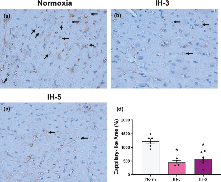
FIGURE 4.

Capillary density. CD31/PECAM staining in heart sections of mice under hypoxia intermittent. Representative microphotographs were obtained using a 40x objective (a, b, c). Arrows indicate the CD‐31/PECAM stained in the left ventricle. (d) *different from normoxia (p < 0.001). N = 4–6/group. Data mean ±SEM. Scale bar: 50 μm
