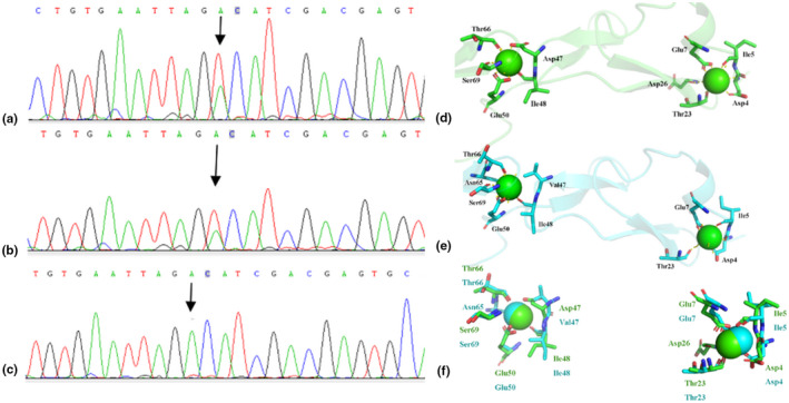
FIGURE 3.

(a) DNA sequencing revealed a heterozygous sing‐base (c.3344A>T, NM_001999.4) affected patients (I1, II2, II6, III1, and III3); (b)fetal amniotic fluid DNA revealed the existence of this mutation;(c) unaffected people (I2, II1, II3, II4, II5, II7, and III2 in this family do not have this mutation. (d) Wild type modeled structure of cbEGF domains 12–13 of the fibrillin‐2, the blue ball means Ca2+. (e) Mutant type modeled structure of cbEGF domains 12–13 of the fibrillin‐2, Val 1115 the blue ball means Ca2+. The protein modeling is achieved by PyMOL Molecular Graphics System (Version 2.3.0) according to FBN1 structural coding (Smallridge et al., 2003). (f) Compare wild type modeled structure of cbEGF domains 12–13 of the fibrillin‐2 and mutant type modeled.
