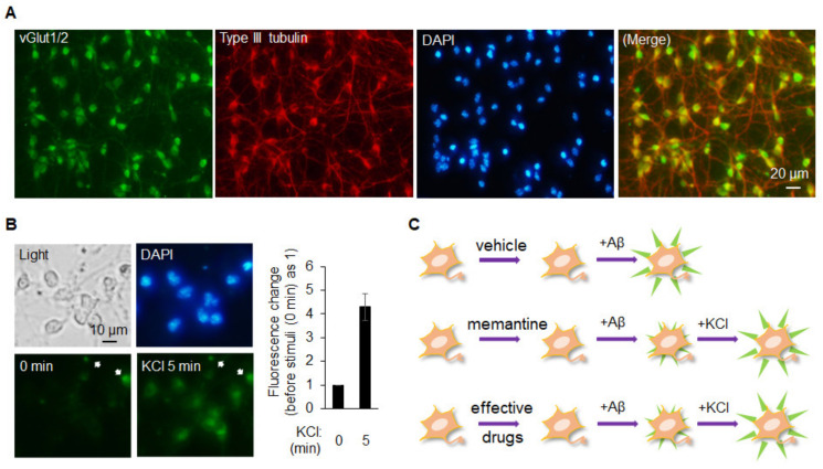
Figure 3.
Glutamatergic neurons differentiated from ESCs could provide an alternative resource for establishing the drug screening system/platform: (A) neurons differentiated from mESCs for around 7 days were fixed and immunocytochemistry was performed using vGLUT1/2 or type III tubulin as the primary antibody. A merged image of vGLUT1/2 (green) and type III tubulin (red) is shown; (B) neurons differentiated from mESCs were incubated with 1 µM DiBAC4(3), a slow-response voltage sensitive fluorescent dye, for 30 min in KRH solution prior to the experiment. The fluorescent intensity was monitored and recorded every minute after 45 mM KCl was added to the solution. Only the images before (0 min) and after (5 min) the stimulation are shown here. The arrows indicate the nonspecific signals which are used as controls for fluorescent intensity. Intensity changes were quantified, plotted and are shown on the right panel. Data are shown as means ± SD; (C) a cartoon is drawn schematically to indicate the use of these neurons for drug screening platform. Top panel, Aβ1-42-induced abnormal depolarization will be observed. Middle panel, memantine, a drug used as part of the clinical treatment of AD patients, shall be able to block the Aβ1-42-induced abnormal depolarization. At the end of experiment, these neurons shall be stimulated with KCl to confirm their responsiveness. The use of memantine functions as a positive control for the platform. Bottom panel, the effective drugs can be screened by using the same logic with the one in memantine.

