Figure 2.
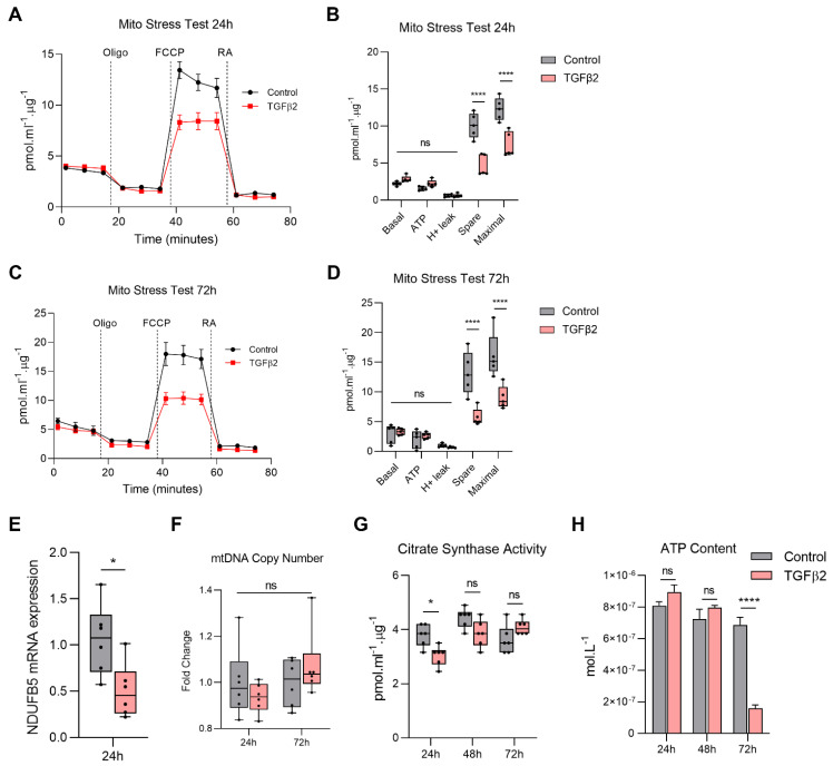
TGFβ2 reduces mitochondrial respiration in RPE. Real-time measurement of oxygen consumption rate (OCR) using the Seahorse XF24 BioAnalyzer to assess OXPHOS parameters: basal respiration, ATP-linked respiration, proton leak, spare respiratory capacity and maximal respiration based on responses to drug injections of oligomycin (Oligo), FCCP and rotenone and antimycin A (RA) at (A,B) 24 h and (C,D) 72 h in ARPE-19 treated with and without TGFβ2 (n = 5, one-way ANOVA with Tukey’s post-hoc analysis). (E) Quantification of NDUFB5 gene expression following treatment with or without TGFβ2 at 24 h (n = 6, unpaired t-test). (F) Quantification of mitochondrial DNA (mtDNA) copy number using qPCR with and without TGFβ2 for 24 and 72 h (n = 6, one-way ANOVA with Tukey’s post-hoc analysis). Error bars are means ± SEM. * p ≤ 0.05; ns, not significant. (G) Quantitation of citrate synthase activity (n = 6) and (H) Intracellular ATP content at 24, 48 and 72 h with or without TGFβ2 (n = 3, one-way ANOVA with Tukey’s post-hoc analysis). Error bars are means ± SEM. * p ≤ 0.05; **** p ≤ 0.0001; ns, not significant.

