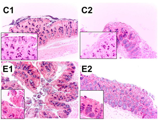
Figure 5.
Histological sections of the colon from each experimental group: C1 wild-type mice fed a standard diet, C2 ApcMin/+ fed a standard diet, E1 ApcMin/+ fed a standard diet supplemented with microencapsulated Bf and Lg, E2 ApcMin/+ fed a standard diet supplemented with microencapsulated Bf, Lg, and quercetin. The images were taken at 20× and at 63× (left lower panel, increased epithelial).

