Figure 1.
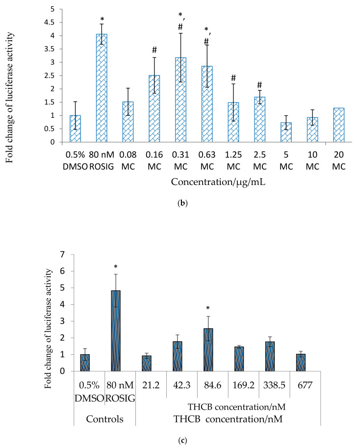
PPARγ ligand screening of (a) Rosiglitazone at concentrations ranging from 0–140 nM, (b) Momordica charantia (MC) fruit crude methanol extract from 0.08–20 μg/mL, (c) 3β,7β,25-trihydroxycucurbita-5,23(E)-dien-19-al (THCB) at concentration from 21.2–677 nM. HepG2 cells were transfected with PPRE-Tk-Luc, pSV-sport PPARγ, pSV-Sport RXRα, and pRL-CMV for overnight and treated as mentioned in the graph respectively. 80 nM rosiglitazone (Rosig) was used as the positive control, while 0.5% DMSO (which was used as carrier to dissolve the compound) was used as the negative control. After 23 h, luciferase activity was determined. Luciferase ratio of the cells treated with 0.5% DMSO was assigned to the value 1, and the results were presented as fold luciferase ratio relative to this value. Statistical analysis was conducted using one-way ANOVA (p < 0.05) where ‘*’ denotes statistically significant difference as compared to untreated control, while ‘#’ denotes not statistically different when compared to positive control rosiglitazone where applicable.


