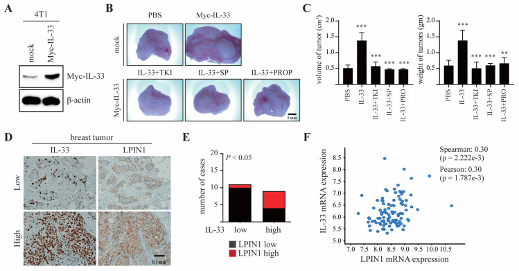
Figure 6.
LPIN1 overexpression is associated with breast mammary tumorigenesis. (A) Expression levels of IL-33 in 4T1/mock and 4T1/Myc-IL-33. (B,C) Myc-IL-33-overexpressing 4T1 cells were treated with 100 μM TKI or 50 μM SP600125 or 0.5 mM propranolol. The treated cells were injected into the mammary glands of BALB/c mice and allowed to grow until tumors formed (14 days). Representative pictures of tumors (B) and tumor volume and weights (C) are shown. Columns, mean of triplicate samples. bars, S.D. **, p < 0.01, ***, p < 0.001. (D,E) Representative samples showing results of immunohistochemical analysis of breast-infiltrating duct carcinoma performed with the indicated antibodies on adjacent sections of the samples. In each sample, IL-33 and LPIN1 levels were semi-quantified in a double-blind manner as high or low, according to the standards presented and statistically analyzed in (E). Their correlation was analyzed using Fisher’s exact test (p < 0.05). (F) The correlation between IL-33 and LPIN1 mRNA expression was assessed using expression data from Molecular Taxonomy of Breast Cancer International Consortium (METABRIC) and TCGA (n = 6).

