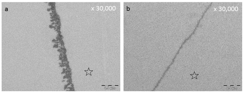
Figure 5.
Both TEM images visualize the 2 h pellicle after a pellicle formation time of 1 min and rinsing with tannic acid for 10 min (a,b). (b) Sample generated after an additional treatment with HCl (pH 2.3) for 1 min in vitro (Hertel et al. (2017) [177]. In comparison, Figure 7a shows a 2 h control pellicle without rinsing. The former enamel site is marked with an asterisk.

