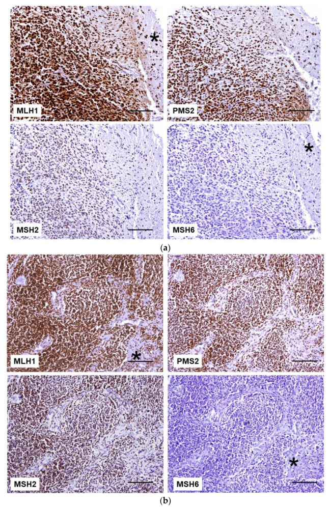
Figure 2.
MMR protein IHC in brain tumors. (a) Glioblastoma cells and normal brain tissue (*) retained the expression of MLH1, PMS2, and MSH2 proteins. MSH6 staining is lost in both tumor cells and normal tissue (*). (b) In malignant astrocytoma, neoplastic, stromal, and endothelial cells are immunoreactive to anti-MLH1 (*), anti-PMS2, and anti-MSH2 antibodies. In contrast, the lack of MSH6 staining is observed in all cells (*). Scale bar: 100 µm.

