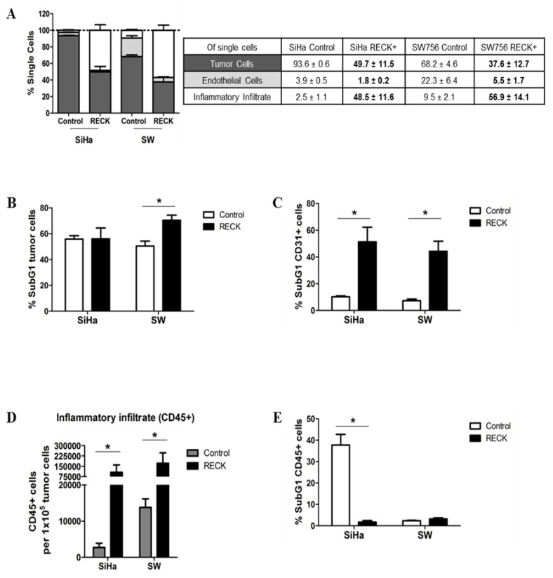
Figure 3.
RECK over expression is associated with alterations in tumor microenvironment content. The injection protocol for tumor establishment was the same as described in Figure 1A. Here, the experiment ended once most of the animals from the control group presented tumors with 500 mm3. (A). Intra-tumoral populations were characterized by immunostaining with anti-CD31 and CD45 antibodies, followed by flow cytometry. Tumor fractions of at least three animals from each group were used for the analysis and at least 1 × 10⁵ cells/events were acquired. The EGFP+ population was identified as tumor cells. CD31+ and CD45+ populations identified endothelial cells and inflammatory infiltrate, respectively. Significant differences in population proportion are indicated in bold font in the table. (B,C,E) Tumor (B), endothelial (C) and inflammatory (E) cell populations stained with DAPI were evaluated on hypodiploid DNA content (SubG1) using flow cytometry. (D) Local inflammatory infiltrate population relative to 100,000 tumor cells. * p < 0.05 by Kruskal–Wallis (3A) or Mann–Whitney (3B–E) test.

