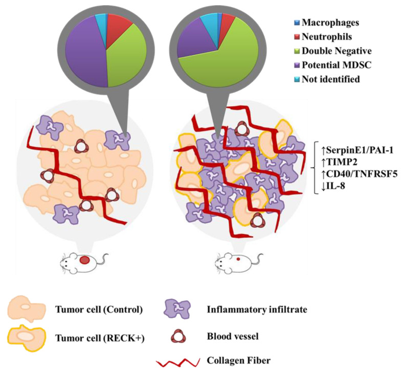
Figure 8.
Proposed global effects of RECK expression in a cervical cancer model. Animals s.c. injected with cervical cancer derived cells transduced to over express RECK present delayed establishment and reduced tumor volume when compared to control. RECK over expressing cells are represented with light orange cell membrane and the red circles in mice lateral side represent the induced tumors. RECK+ tumors also showed reduced proportion of tumor and endothelial cells, the latter was depicted here in dark red blood vessels. Moreover, we observed specific increased proportion of Double negative inflammatory infiltrate (pie charts in the top) and higher collagen content (bright red linear structures) in RECK+ tumors. Finally, RECK over expression correlated with higher levels of SerpinE1/PAI-1, TIMP-2 and CD40/TNFRSF5 proteins, while presented an inverse correlation with IL-8 protein expression (on the far-right side).

