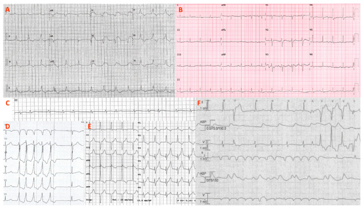
Figure 1.
ECG and arrhythmias. Representative ECGs and arrhythmias from the patient series (P1–P7) are shown. (A) Atrioventricular dissociation with huge anterior ST elevation (P7); (B) sinus tachycardia with low QRS voltages and diffuse repolarization abnormalities (P2); (C) paroxysmal atrial fibrillation detected by ICU telemonitoring (P7); (D) nonsustained ventricular tachycardia (P4); (E) transitory, self-limited, accelerated junctional rhythm (P1); (F) polymorphic and irregular nonsustained ventricular tachycardias during ICU stay (P5). ICU = intensive care unit.

