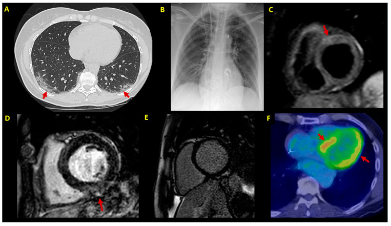
Figure 2.
Imaging findings. Imaging findings at patient (P1–P7) diagnostic workup. (A) Chest CT scan showing bilateral patchy ground-glass opacities (arrows) (P3); (B) chest X-ray in a patient (P7) with cardiogenic shock supported by IABP, VA-ECMO and temporary pacemaker; (C) CMR in a patient with infarct-like acute myocarditis associated with COVID-19 (P3); T2-STIR sequence shows edema in the anterior basal segment (arrow); (D) LGE sequences in a patient (P5) showing mild inferior mid-myocardial/subepicardial LGE (arrow); (E) absence of LGE by 3-month follow-up CMR in a patient (P4) with fulminant myocarditis at presentation; (F) 3-month follow-up FDG-PET scan in an ICD carrier (P5) with virus-negative myocarditis; abnormal left ventricular FDG uptake is shown (arrows). CMR = cardiac magnetic resonance; CT = computed tomography; EMB = endomyocardial biopsy; FDG-PET= 18F-fluorodeoxyglucose positron emission tomography; IABP = intra-aortic balloon pump; ICD = implantable cardioverter defibrillator; LGE = late gadolinium enhancement; LVEF = left ventricular ejection fraction; STIR = short-tau inversion recovery; VA-ECMO = venoarterial extracorporeal membrane oxygenator.

