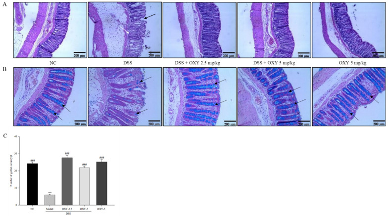
Figure 3.
Effect of OXY on histopathological changes in the distal colon (n = 8/group). The middle part of distal colons was sectioned and stained with hematoxylin and eosin (H&E) (A), infiltrated inflammatory cells (white arrow) and damaged crypt structure (black arrow) as well as with Alcian blue (B), goblet cell (black arrow). Goblet cells within a crypt were counted (C). NC, normal control group; model, DSS-induced colitis model group; DSS + OXY-2.5, DSS-induced colitis rats treated with OXY (2.5 mg/kg) group; DSS + OXY-5, DSS-induced colitis rats treated with OXY (5 mg/kg) group; OXY-5, rats treated with OXY (5 mg/kg) alone group. The values are expressed as mean ± SEM. Significant differences among groups were evaluated via ANOVA with Tukey’s post hoc HSD. *** p < 0.001 compared with the NC group; ### p < 0.001 compared with the model group.

