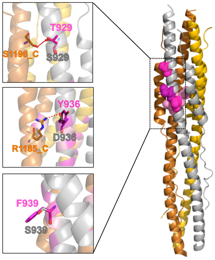
Figure 4.
Mutants in the post-fusion conformation. Right: Cartoon representation of the SARS-CoV-2 S protein in its post-fusion trimeric conformation (the three monomers are colored in silver, gold, and copper, PDB ID: 6LXT). The color code is the same in Figure 3. Mutations in the HR1 fusion core are shown in a “dots” representation for chain A. Left: Focus on the structural context of each wild-type residue (silver sticks) and corresponding mutant (purple sticks). H-bonds are shown as red, dashed lines.

