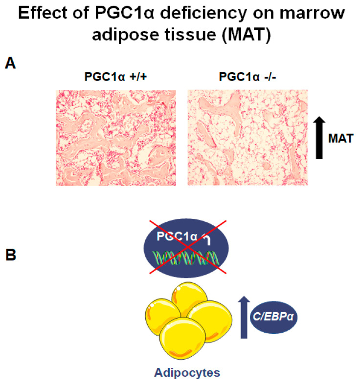
Figure 2.
PGC1α deletion affects marrow adipose tissue (MAT). (A) Photomicrographs of hematoxylin and eosin-stained sections of MAT from PGC1α +/+ and PGC1α −/− (magnification: 20×) show an increased number of adipocytes in the absence of PGC1α (unpublished data). (B) Schematic representation of the effect of PGC1α deletion in bone marrow adipocytes consisting of the increase of C/EBPα expression.

