Figure 1.
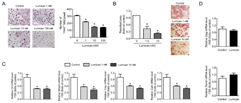
Lumican suppresses osteoclast differentiation and in vitro bone resorption. (A) Primary mouse BMMs were cultured for 4 days with 30 ng/mL RANKL, 30 ng/mL M-CSF, and the indicated concentrations of lumican. After the cells were stained with TRAP, TRAP-positive multinucleated cells (MNCs) (≥3 nuclei/cell) were counted to assess osteoclast differentiation (n = 5). (B) Mouse BMMs were cultured on dentin slices for 10 days in the presence of 30 ng/mL RANKL, 30 ng/mL M-CSF, and 1 nM or 10 nM lumican and stained with hematoxylin to visualize pit formation (n = 3). (C) Quantitative RT-PCR analysis of osteoclast differentiation markers in BMMs exposed to 30 ng/mL RANKL, 30 ng/mL M-CSF, and 1 or 10 nM lumican for 4 days (n = 4). (D) Quantitative RT-PCR analysis to measure Opg and Rankl expression in mouse calvaria osteoblasts exposed for 7 days to 10 mM β-glycerophosphate and 50 mg/mL ascorbic acid, with or without 10 nM lumican (n = 3). Scale bars: 100 μm (A) and 50 μm (B). Data are means ± SEM. * p < 0.05 vs. untreated control.

