Figure 3.
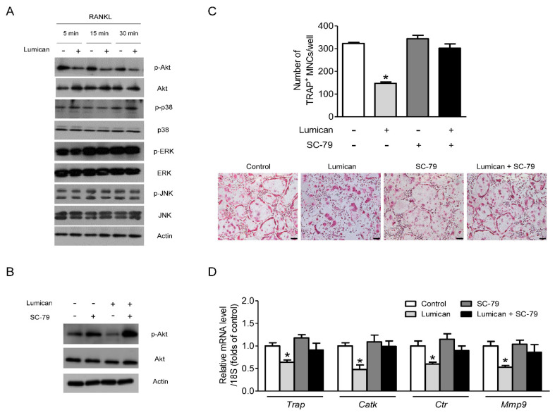
Suppression of the Akt signal mediates the effects of lumican on osteoclastogenesis. (A) Western blot to assess RANKL-dependent phosphorylation of downstream kinases in mouse BMMs after exposure to 10 nM lumican and 30 ng/mL RANKL following culture for 3 days in the presence of 30 ng/mL M-CSF (n = 5). (B) Western blot analysis to assess the phosphorylation of Akt in mouse BMMs after exposure to 30 ng/mL RANKL, 10 nM lumican, and 2 μg/mL SC-79 (Akt activator) following culture for 3 days in the presence of 30 ng/mL M-CSF (n = 3). (C,D) Primary mouse BMMs were co-treated with 30 ng/mL RANKL, 30 ng/mL M-CSF, 10 nM lumican, and 2 μg/mL SC-79 for 4 days (n = 4). (C) After the cells were stained with TRAP, the number of TRAP-positive multinucleated cells (MNCs) (≥ 3 nuclei/cell) was determined to assess osteoclast differentiation. (D) Quantitative RT-PCR analysis was performed to determine the levels of osteoclast differentiation markers. Scale bars: 100 μm (C). Data are means ± SEM. * p < 0.05 vs. untreated control.

