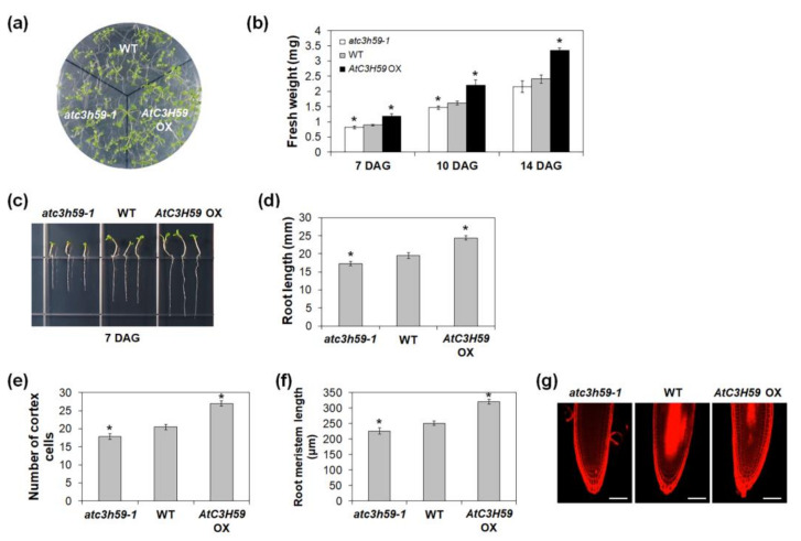
Figure 5.
Seedling development of AtC3H59 OXs and atc3h59 mutants. (a) Fourteen-day-old WT, AtC3H59 OXs, and atc3h59 mutants grown on MS plates under SD conditions. (b) Fresh weights of shoots of WT, AtC3H59 OX, and atc3h59 mutant seedlings grown on MS plates under SD conditions at 7, 10, and 14 DAG. Data shown are the mean ± S.D. (n = 5). (c) Elongation of primary roots of WT, AtC3H59 OXs, and atc3h59 mutants at 7 DAG. (d) Primary root lengths of WT, AtC3H59 OXs, and atc3h59 mutants grown on MS plates under SD conditions were measured at 7 DAG. Data shown are the mean ± S.D. (n = 24). (e) The number of root meristem cortex cells of WT, AtC3H59 OXs, and atc3h59 mutants grown on MS plates under SD conditions were measured at 7 DAG. Data shown are the mean ± S.D. (n = 12). (f) Root meristem length of WT, AtC3H59 OXs, and atc3h59 mutants grown on MS plates under SD conditions were measured at 7 DAG. Data shown are the mean ± S.D. (n = 12). (g) Confocal microscopy of roots of WT, AtC3H59 OXs, and atc3h59 mutants grown on MS plates under SD conditions at 7 DAG. Roots were excised from seedlings, stained with 10 μM propidium iodide, and examined by confocal microscopy. Scale bars represent 50 μm. In (b,d–f), * indicate t-test p < 0.05. At least three biological replicates showed similar results. Three independent T1 lines of AtC3H59 OXs showed very similar results, with one shown here.

