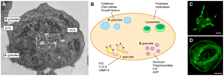
Figure 1.
(A) Electron micrograph of resting platelet. Indicated: open canalicular system (OCS), dense tubular system (DTS), α-granules, δ-granules that contain small molecules. (B) Graphical representation of the main secretory granules of platelets and their contents. Indicated: protein disulfide isomerase (PDI), with toll-like receptor 9 (TLR), vesicle-associated membrane protein 8 (VAMP-8). (C) A platelet forming filopodia and (D) a fully spread platelet. Platelets were stained for F-actin with phalloidin-Alexa Fluor 488, and visualized by a confocal laser scanning microscope.

