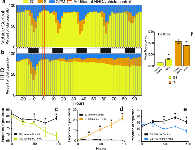
FIG 2.
HHQ triggers stalling in S phase. (a and b) The cell cycle stage was quantified by profiling the fluorescence (575/25 nm), a proxy for DNA content, of propidium iodide-stained E. huxleyi cultures (n = 3) exposed to either the vehicle control (DMSO) (a) or 100 ng ml−1 HHQ (b) for 96 h. (c through e) The proportion of cells in each cell stage was determined from density plots of the distribution of cells with various DNA contents ranging from 2N (G1) to 4N (G2) at T0, T24, T48, T72, and T96. Cells with intermediate DNA content were denoted as S phase, as the genome replicated. Each plot represents the mean ± standard deviation for triplicate samples (P < 0.05 by ANOVAR). (f) Mean fluorescences (575/25 nm) of G1- and S-phase cells treated with the vehicle control (DMSO) or 100 ng ml−1 HHQ for 96 h and stained with propidium iodide were compared via Welch’s approximate t test (P < 0.01). As DNA replication occurs only in S phase, the increase in the mean fluorescence for HHQ-treated cells that fall within the G1 gate suggests that these cells are currently in S phase but stall early in the process of DNA synthesis and are unable to synthesize enough additional DNA to fall within the S-phase region.

