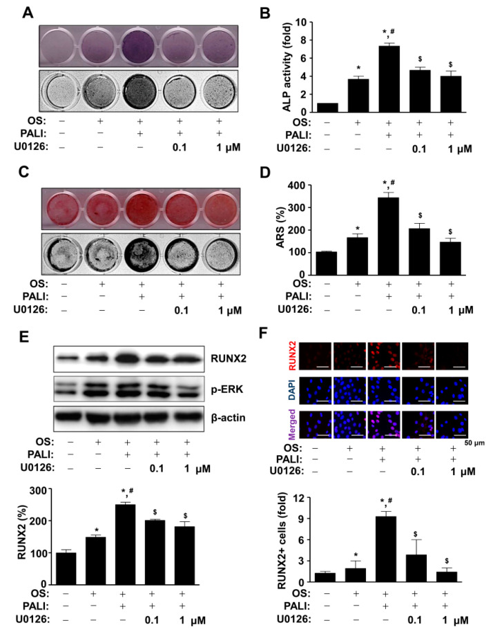
Figure 5.
Effects of paeonolide (PALI)-induced ERK1/2 signals on osteoblast differentiation. (A,B) After pre-osteoblasts were treated with PALI (10 μM) in the presence or absence of U0126 (1 and 10 μM) during osteogenic supplement medium (OS)-induced osteoblast differentiation, alkaline phosphatase (ALP) staining was analyzed using a scanner (upper) and a colorimetric detector (bottom) (A), and the enzymatic activity of (ALP) was measured at 405 nm using a spectrophotometer and is expressed as a bar graph. (C,D) Alizarin red S (ARS) staining was analyzed using a scanner (upper) and a colorimetric detector (bottom) (C), and the intensity of mineralized nodule formation was quantified and is expressed as a bar graph (D). (E,F) Equal amounts of lysates were analyzed using Western blot analysis and detected with antibodies against RUNX2 and phospho-ERK (p-ERK). β-Actin was used as a loading control (E). The fixed cells were analyzed using immunofluorescence analysis and immunostained with antibody against RUNX2 (red). DAPI (blue) was used as a nuclear marker. The bottom panels show the merged images (purple) of the first and second panels. RUNX2+ cells (fold) were expressed as a bar graph. Scale bar: 50 μm. Data are representative of the results of three independent experiments and values are expressed as mean ± SEM. *, p < 0.05 indicates statistically significant difference compared with the control; #, statistically significant difference compared with OS (p < 0.05); $, statistically significant difference compared with the OS + PALI (p < 0.05).

