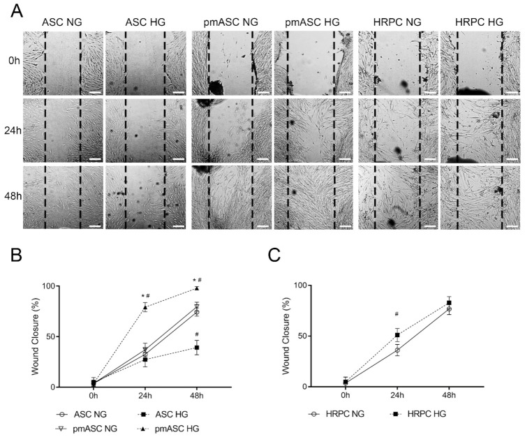
Figure 3.
Cell migration ability evaluated by wound-healing assays in human adipose mesenchymal stem cells cultured in basal medium (ASC) or in pericyte medium (pmASC), and in cultures of human retinal pericytes (HRPC). In some samples of each culture type, glucose was added to the culture medium (High Glucose, 25 mM, HG) whereas other samples were kept in normal glucose (NG) condition. (A): Representative pictures of each sample taken immediately after the scratch (0 h), after 24 h and 48 h of culture. Scale bar: 100µm (Magnification: 4×). Percentage of wound closure was quantified by Image J software for ASC and pm ASC cultures (B), and HRPC cultures (C). Values are expressed as mean ± SEM of three independent experiments. * p < 0.05 pmASC vs. ASC; # p < 0.05 HG vs. NG. Two-way ANOVA, followed by Sidak’s test.

