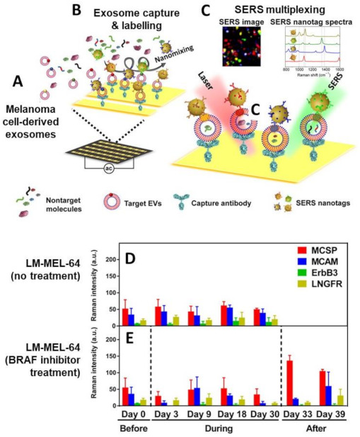
Figure 14.

(A–C) Schematic of the exosome phenotyping using SERS-encoded nanoparticles: (A) Exosomes are secreted by melanoma cells with a BRAF V600E mutation in the culture medium or into circulation; (B) The exosome containing sample is injected, together with SERS tags, into the nanomixing chip equipped with capture antibodies; (C) Upon removal of non-target molecules (e.g., protein aggregates and apoptotic bodies) and unbound SERS tags, SERS mapping is performed to provide the SERS phenotyping of the captured exosomes. The false-color SERS image is generated from the characteristic peak intensities of each SERS tags (MCSP-MBA, red; MCAM-TFMBA, blue; ErbB3-DTNB, green; LNGFR-MPY, yellow). (D,E) Phenotypic alterations of exosomes derived from melanoma patient-derived LM-MEL-64 cell line in response to BRAF inhibitor treatment at different times (before, during and after treatment). Anti-CD63 antibodies were used in the capturing area. Adapted with permission from [99]. © The Authors, some rights reserved; exclusive licensee AAAS. Distributed under a Creative Commons Attribution NonCommercial License 4.0 (CC BY-NC). Available online: http://creativecommons.org/licenses/by-nc/4.0/ (accessed on 30 April 2021).
