Figure 2.
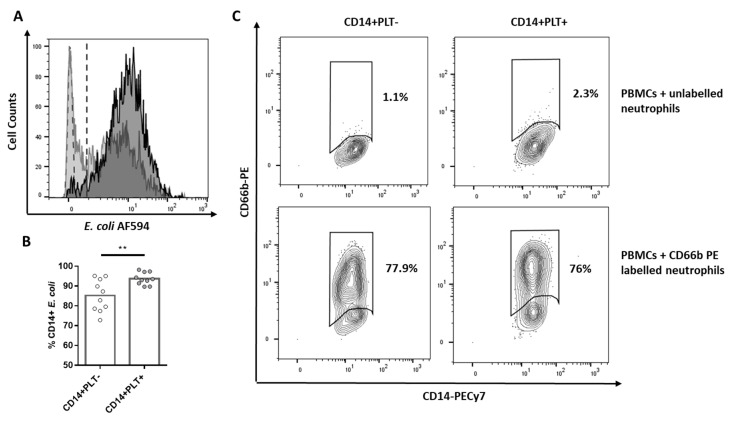
E. coli phagocytosis of HD monocytes with or without bound platelets. PBMCs were cultured with E. coli bioparticles stained with Alexa Fluor 594. (A) The representative overlapping histogram plot is shown with the phagocytosis of CD14+PLT- in light gray and of CD14+PLT+ in dark gray. PBMCs without E. coli (light green dotted histogram) are the control to set up threshold of negative cells. Dotted line indicates positivity threshold. (B) Phagocytosis of E. coli by CD14+PLT- and CD14+PLT+ from 10 independent experiments. (C) Phagocytosis of apoptotic neutrophils (efferocytosis). A representative experiment is shown (n = 8) from 6 independent experiments. Data are expressed as percentages of gated monocytes. The statistical analysis was performed using the paired t-test. ** p < 0.01.

