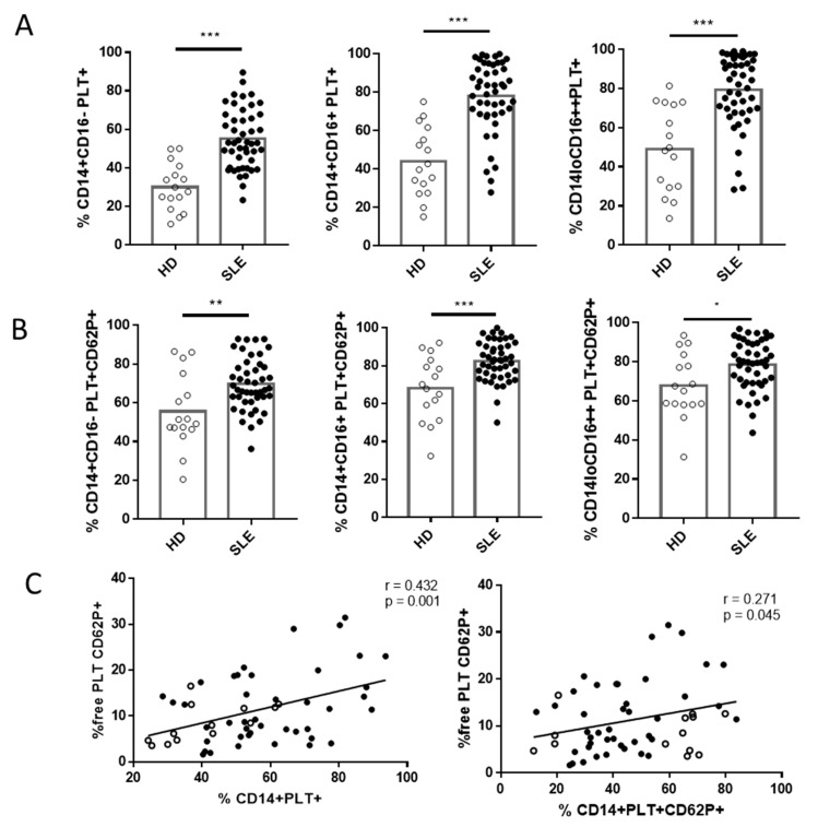
Figure 4.
Monocytes with bound platelets and their relationship with free platelets in HD and SLE patients. (A) Percentage of monocyte subsets with bound platelets (PLT+) in HD and SLE patients. (B) Percentage of monocyte subsets with activated bound platelets (PLT+CD62P+) in HD and SLE patients. (C) Correlation between the percentage of CD14+PLT+ and CD14+PLT+CD62P+ with the percentage of free activated platelets (free PLT CD62P+) in HD and SLE patients. White circles represent HD (n = 16), and black circles represent SLE patients (n = 49). Statistical analysis was performed using the unpaired t-test (CD14+PLT+, CD14+PLT+CD62P+) for comparisons between HD and SLE and Spearman’s correlation for correlation analysis. * p < 0.05, ** p < 0.01, and *** p <0.001.

