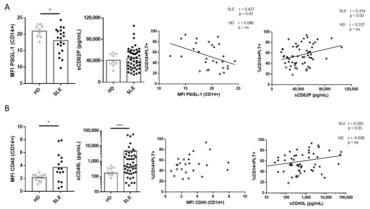
Figure 5.
Expression of PSGL-1 and CD40 on monocytes and quantification of soluble CD62P (sCD62P) and CD40L (sCD40L) in plasma from HD and SLE patients and their association with the percentage of monocytes with bound platelets (CD14+PLT+). (A) PSGL-1 gMFI of monocytes from HD (n = 9) and SLE patients (n = 20). Levels of sCD62P in plasma from HD (n = 9) and SLE patients (n = 49). PSGL-1 and sCD62P correlations with the percentage of CD14+PLT+. (B) CD40 MFI of monocytes from HD (n = 12) and SLE patients (n = 15). Levels of sCD40L in plasma from HD (n = 9) and SLE patients (n = 49). CD40 and sCD40L correlations with the percentage of CD14+PLT+. White circles represent HD, and black circles represent SLE patients. Statistical analysis was performed using the Mann–Whitney test for comparisons between HD and SLE and Pearson’s (PSGL-1 and sCD62P) or Spearman’s (CD40 and sCD40L) correlation for correlation analysis. * p < 0.05, *** p < 0.001.

